Cardiac Electro Physiology in Raipur – Heart Rhythm Clinic

Cardiac Treatments

R.F. Ablation

Radiofrequency ablation (also known as fulguration) is a medical procedure that involves the heating of medium frequency alternating current to ablate a part of the electrical conduction systems of the heart, tumour, or other dysfunctional tissue.

SVT Ablation

SVT (Supraventricular Tachycardia), also known as ablation, uses heat or cold energy in order to create tiny scars in your heart to block any abnormal electrical signals and return to a normal rhythm.

A.F. Ablation

The atrial fibrillation ablation is usually done with thin, flexible catheters inserted through the veins and arteries to the heart. Ablation can also be performed during cardiac surgery, although it is less common.

V.T. Ablation

Ventricular Tachycardia is a condition in which the electrical signals from your heart cause your lower heart chambers (ventricles) to beat too fast.

W.P.W. Syndrome

Wolff-Parkinson-White syndrome is a medical condition in which an extra electrical pathway in the heart leads to periods of rapid heart rate (tachycardia).

3D Mapping & Ablation

3D mapping allows for a multidimensional view of the anatomy of the heart. Your doctor will stimulate the heart muscle during cardiac mapping to monitor its rhythm and identify irregularities (cardiac arrhythmia).

Angiography and Angioplasty

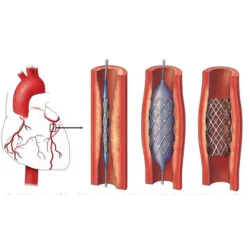
Angiography is used to examine the blood vessels and determine if there are any potential heart conditions. However, angioplasty uses narrowed arteries to treat the condition.

Pacemaker Implantation

A pacemaker insertion is the instalment of a small electronic device. It is typically placed just below the collarbone in the chest to regulate slow electrical problems with your heart.

CRT-P Implantation

C.R.T. has been clinically proven to work as a treatment for heart failure patients. The C.R.T. device sends small electrical impulses to the lower chambers of the heart to cause them to beat more efficiently.

I.C.D. Implantation

A cardioverter-defibrillator (I.C.D.) which can be implanted is a small battery-powered device placed in your chest to detect and stop abnormal heartbeats (arrhythmias).

Genetic Arrhythmia

It is also called familial arrhythmia when the condition is passed down from one generation to another. Genetic arrhythmia syndrome symptoms can vary depending upon the cause and type of arrhythmia.

Conduction System Pacing

It is a pacing method that involves the implantation of permanent pacing leads at different sites in the cardiac conduction network. It also includes His bundle branch pacing and left-bundle branch pacing.

Heart Failure

Congestive heart disease, commonly known as heart failure, happens to be a condition in which the heart muscle fails to pump blood as efficiently. This can lead to blood backup, fluid buildup in the lungs, and shortness of breath.

Syncope

Syncope, pronounced "sin ko pea," is the medical term that refers to fainting or falling asleep. A temporary decrease in blood flow to the brain is what causes syncope.

Echo Cardiography

An echocardiogram (or echo) is a graphic representation of the heart's movements. An echo test uses high-frequency sound waves (high-frequency ultrasound) to take pictures of your heart.

Treadmill Test

To determine the effect of exercise on the heart, a treadmill exercise stress test is used. Doctors can detect irregular heart rhythms (arrhythmias) and diagnose coronary artery disease.

E.C.G. (Electro Cardiography)

An electrocardiogram is an electrocardiogram, also known as EKG or E.C.G. An electrogram of your heart is a graph that shows voltage and time of electrical activity. It's done using electrodes on the skin.

Holter Monitoring

The Holter monitor is a portable electrocardiogram (ECG). While you're away from your doctor's office, it continuously records the electrical activity in the heat for 24 hours or more. The standard, or "resting," E.C.G. is the most common test to evaluate the heart.

Cardiac Imaging

Cardiac imaging is a subspecialty in diagnostic radiology. A cardiac radiologist is responsible for interpreting medical images and performing diagnostics to determine the severity of heart diseases. This includes leaky valves, heart disease, and other defects.

 
\